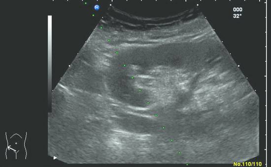
Ultrasound Image Obtained During Percutaneous Renal Biopsy Arrow

Ultrasound Image Obtained During Percutaneous Renal Biopsy Arrow Ultrasound image obtained during percutaneous renal biopsy. arrow showing the needle, characterized by linear hyperechoic imaging, entering the renal parenchyma at the lower pole. Ultrasound image obtained during biopsy shows 18 gauge needle tip (black arrow) in center of mass (white arrow). biopsy results confirmed that mass is fat poor aml, and unnecessary treatment and its associated risks were avoided.

Ultrasound Image Obtained During Percutaneous Renal Biopsy Arrow Figure: ultrasound guided core needle biopsy of a native kidney. a. color doppler shows relatively decreased vascularity of the lower pole. b. the arrow shows a sample being obtained from the renal cortex. Percutaneous renal biopsy, utilizing either ultrasound or ct, allows for an accurate, reliable method of acquiring renal tissue for histopathological assessment. Ultrasound image obtained during percutaneous renal biopsy. arrow showing the needle, characterized by linear hyperechoic imaging, entering the renal parenchyma at the lower pole. The following short article is intended to show how to perform a renal biopsy and to demonstrate the traditional ward biopsy and the “radiological” method using an electronic guide system.

Percutaneous Renal Biopsy Radiology Key Ultrasound image obtained during percutaneous renal biopsy. arrow showing the needle, characterized by linear hyperechoic imaging, entering the renal parenchyma at the lower pole. The following short article is intended to show how to perform a renal biopsy and to demonstrate the traditional ward biopsy and the “radiological” method using an electronic guide system. Real time ultrasound guidance (rt usg) is currently considered the “standard of care” and is the predominant imaging modality reported for percutaneous native renal biopsy. Ultrasound image obtained during percutaneous renal biopsy. arrow showing the needle, characterized by linear hyperechoic imaging, entering the renal parenchyma at the lower pole. Current biopsy techniques involve ct or ultrasound guidance with small gauge needles. the risks of renal biopsy are minimal. the purpose of this article is to discuss the history of, indications and rationale for, and approach to imaging guided percutaneous renal biopsies. We conducted a retrospective study over a seven year period to evaluate the incidence and characteristics of complications following real time ultrasound guided percutaneous kidney biopsy at a single tertiary center.

Percutaneous Renal Biopsy Radiology Key Real time ultrasound guidance (rt usg) is currently considered the “standard of care” and is the predominant imaging modality reported for percutaneous native renal biopsy. Ultrasound image obtained during percutaneous renal biopsy. arrow showing the needle, characterized by linear hyperechoic imaging, entering the renal parenchyma at the lower pole. Current biopsy techniques involve ct or ultrasound guidance with small gauge needles. the risks of renal biopsy are minimal. the purpose of this article is to discuss the history of, indications and rationale for, and approach to imaging guided percutaneous renal biopsies. We conducted a retrospective study over a seven year period to evaluate the incidence and characteristics of complications following real time ultrasound guided percutaneous kidney biopsy at a single tertiary center.
Comments are closed.