Solved Label The Structures On The Following Diagram And Chegg

Solved Label The Structures On The Following Diagram And Chegg Use your free solution no credit card required see answer science biology biology questions and answers label the structures on the following diagram and state the function of each structures. Study with quizlet and memorize flashcards containing terms like label the structures of this prokaryotic cell, label the structures of this eukaryotic cell, which of the following is not a basic process of life? and more.

Solved Label The Following Structures Chegg In the above diagram, the labelling is a follows: 1.) pons 2.) medulla oblongata 3.) brain stem 4.) cerebellum 5.) pre central gyrus 6.) post central gyrus 7.) central sulcus 8.) temporal lobe in the above diagram, the labelling is a follows: 1.) right hemisphere 2.) left hemisphere 3.) longitudinal fissure 4.) frontal lobe 5.) parietal lobe 6. Label the following diagram of an animal cell. include the 15 structures and organelles listed above. your solution’s ready to go! enhanced with ai, our expert help has broken down your problem into an easy to learn solution you can count on. question: label the following diagram of an animal cell. Drag the labels onto the diagram to identify the components of the pulmonary circuit. excess plasma lipids in the form of cholesterol contribute to the formation of atherosclerotic plaques within blood vessel walls. The lower lobe of the left lung is positioned inferiorly (near the diaphragm) and would be closer to label #8. b. middle lobe: the middle lobe is only present in the right lung and lies below the superior lobe but above the inferior lobe. c. superior upper right lobe: label #1 is at the top part of the right lung.

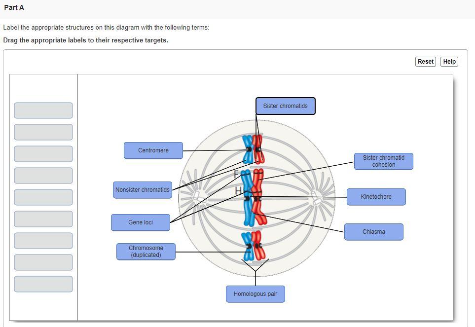
Part A Label The Appropriate Structures On This Chegg Drag the labels onto the diagram to identify the components of the pulmonary circuit. excess plasma lipids in the form of cholesterol contribute to the formation of atherosclerotic plaques within blood vessel walls. The lower lobe of the left lung is positioned inferiorly (near the diaphragm) and would be closer to label #8. b. middle lobe: the middle lobe is only present in the right lung and lies below the superior lobe but above the inferior lobe. c. superior upper right lobe: label #1 is at the top part of the right lung. For the diagram to the right, label the following (1 pt each): a. the name of the organelle b. the name for the entire stack of membranes that is circled c. How is the elisa test quantified? the reaction of a substrate with the enzyme to produce a colored product, thus indicating a positive reaction. label the lymph nodes based on their region or location. Look at the diagram and match each label from the list provided to its corresponding structure. This structure resembles adenosine triphosphate (atp) or a related compound, but with a unique thiol ( sh) group and an amino acid like chain. let's systematically address each part of the question (total: 16 points) by annotating the structure as indicated. 1. (a) circle the functional groups linkages and name them.
Comments are closed.