Solved Example Let Us Divide 0 5 By 0 1 0 01 And 0 001 Learning
Solved Example Let Us Divide 0 5 By 0 1 0 01 And 0 001 Learning Plaque volumes were measured ex vivo in pig carotid and femoral artery specimens by 3 dimensional vascular ultrasound (3dvus) using a 3d matrix (electronic sweep) transducer and its associated 3d plaque quantification software, and were compared with gold standard histology. Programmable, ultrafast ultrasound scanners with a high channel count provide an unprecedented opportunity to optimize volumetric acquisition. in this work, we implement nonlinear processing and develop distributed beamformation to achieve fast acquisition over a 47 centimeter aperture.

Solved Let A 1 0 0 0 0 0 5 0 0 0 0 0 1 3 1 1 3 0 3 Chegg An ex vivo phantom using porcine arteries was used to evaluate the accuracy with which (i) b mode ultrasound, (ii) 3 d tomographic ultrasound (tus), (iii) computed tomography (ct) and (iv) magnetic resonance imaging (mri) measured length, diameters and volume. In this paper, we developed an ultrasound guided focused ultrasound (usgfus) system for low cost 3d volumetric histotripsy using nds. this setup is composed of a rotating imaging. In this study, volumetric ultrasound imaging was performed in the craniotomized rat brain to yield 3d ulm in vivo within 7.5 min of acquisition with a commercial system. Both of these demonstrations were performed in ex vivo tissues infused with sono ink. if optimized and demonstrated in live animal models, these approaches could be a promising way to convert open surgeries to less invasive ultrasound based treatments.

Solved Let A 1 0 0 0 4 0 0 0 5 1 0 0 4 5 0 0 3 0 1 Chegg In this study, volumetric ultrasound imaging was performed in the craniotomized rat brain to yield 3d ulm in vivo within 7.5 min of acquisition with a commercial system. Both of these demonstrations were performed in ex vivo tissues infused with sono ink. if optimized and demonstrated in live animal models, these approaches could be a promising way to convert open surgeries to less invasive ultrasound based treatments. Among the various types of transducers used to obtain three dimensional ultrasound images, this paper focuses on the most standardized probe, the linear array transducer, and provides an overview of the system implementations, imaging results, and applications of volumetric ultrasound imaging from the perspective of scanning methods. These vascular tissue properties can be assessed by recent advances in ultrasound technology, including 3d ultrasound imaging, contrast enhanced ultrasound (ceus), as well as strain elastography (se) and shear wave elastography (swe). This manuscript aims to offer a comprehensive review of 3d srus imaging, clarifying methodologies, clinical applications, and notable challenges that could motivate future research and help facilitate clinical translation. In this article, previous and the latest work on designing a real time or near real time 3d ultrasound imaging system are reviewed. specifically, the data acquisition techniques, reconstruction algorithms, volume rendering methods, and clinical applications are presented.

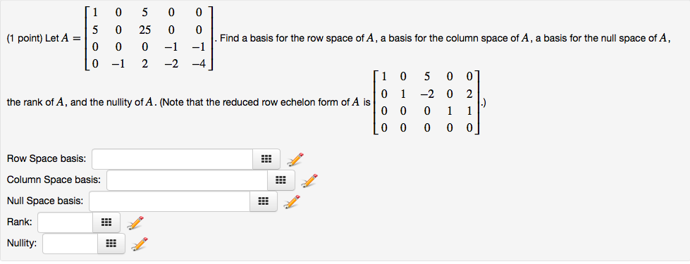
Solved Let A 1 0 5 0 0 5 0 25 0 0 0 0 0 1 1 0 1 2 Chegg Among the various types of transducers used to obtain three dimensional ultrasound images, this paper focuses on the most standardized probe, the linear array transducer, and provides an overview of the system implementations, imaging results, and applications of volumetric ultrasound imaging from the perspective of scanning methods. These vascular tissue properties can be assessed by recent advances in ultrasound technology, including 3d ultrasound imaging, contrast enhanced ultrasound (ceus), as well as strain elastography (se) and shear wave elastography (swe). This manuscript aims to offer a comprehensive review of 3d srus imaging, clarifying methodologies, clinical applications, and notable challenges that could motivate future research and help facilitate clinical translation. In this article, previous and the latest work on designing a real time or near real time 3d ultrasound imaging system are reviewed. specifically, the data acquisition techniques, reconstruction algorithms, volume rendering methods, and clinical applications are presented.
Comments are closed.