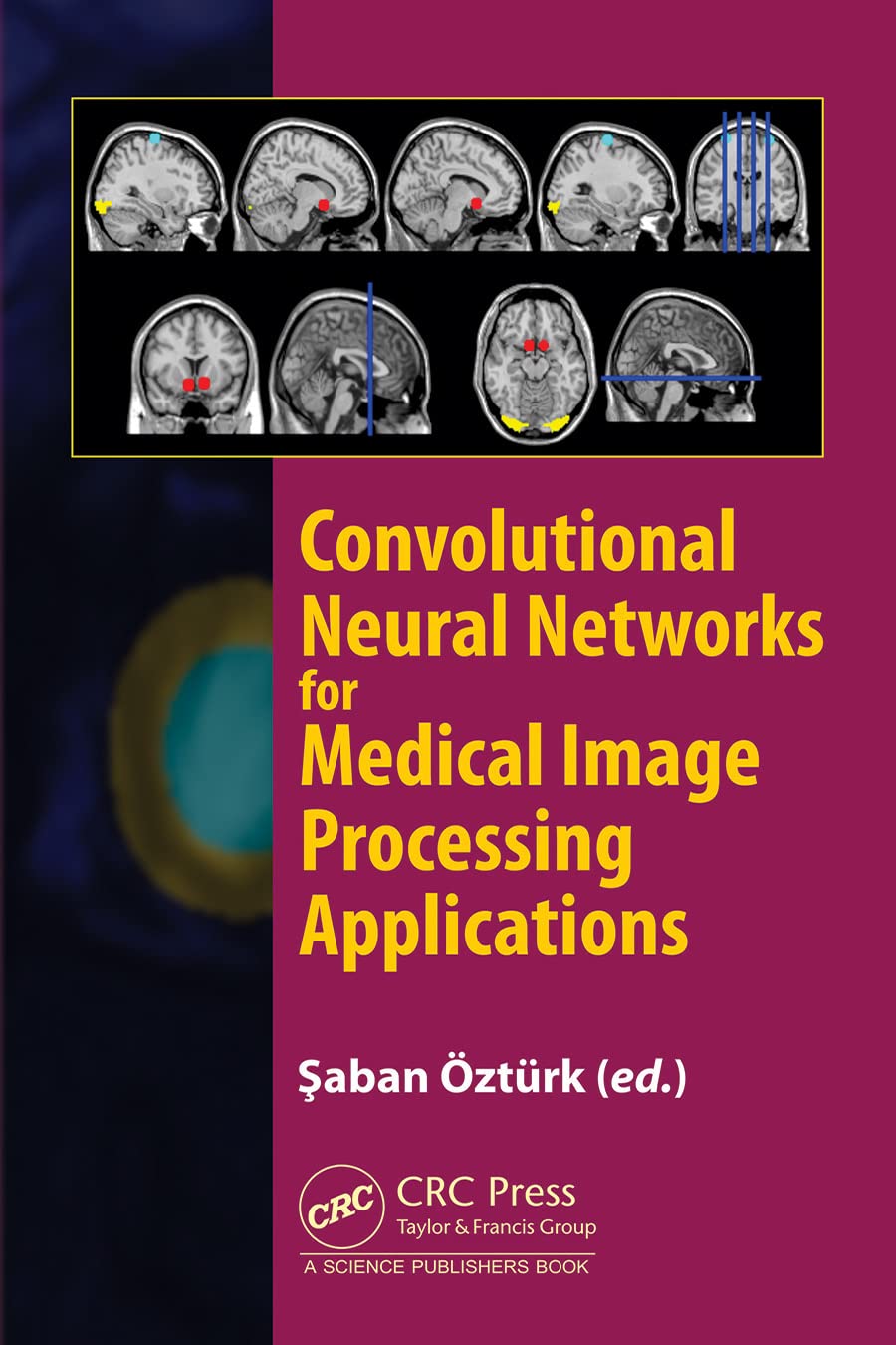
Convolutional Neural Networks For Medical Image Segmentation Paper And

Convolutional Neural Networks For Medical Image Segmentation Paper And In this article, we look into some essential aspects of convolutional neural networks (cnns) with the focus on medical image segmentation. first, we discuss the cnn architecture, thereby highlighting the spatial origin of the data, voxel wise classification and the receptive field. In this paper, we present a network and training strategy that relies on the strong use of data augmentation to use the available annotated samples more efficiently. the architecture consists of a contracting path to capture context and a symmetric expanding path that enables precise localization.

Figure 6 From Medical Image Segmentation Used Unsupervised Convolutional neural networks (cnns) are effective tools for image understanding. they have outperformed human experts in many image understanding tasks. this article aims to provide a comprehensive survey of applications of cnns in medical image understanding. Deep learning, particularly convolutional neural networks (cnns), has significantly advanced segmentation accuracy and efficiency. with the introduction of 3d imaging, research has evolved. This paper provides a progressed convolutional neural community (cnn) version for medical picture segmentation. it specializes in improving the accuracy and speed of today's segmenting of scientific photos and improving modern semantic segmentation of first rate present day clinical photos. In this work we propose an approach to 3d image segmentation based on a volumetric, fully convolutional, neural network. our cnn is trained end to end on mri volumes depicting prostate, and learns to predict segmentation for the whole volume at once.

Convolutional Neural Networks For Medical Image Processing Applications This paper provides a progressed convolutional neural community (cnn) version for medical picture segmentation. it specializes in improving the accuracy and speed of today's segmenting of scientific photos and improving modern semantic segmentation of first rate present day clinical photos. In this work we propose an approach to 3d image segmentation based on a volumetric, fully convolutional, neural network. our cnn is trained end to end on mri volumes depicting prostate, and learns to predict segmentation for the whole volume at once. This work presents a review of the literature in the field of medical image segmentation employing deep convolutional neural networks. the paper examines the various widely used medical image datasets, the different metrics used for evaluating the segmentation tasks, and performances of different cnn based networks. The enhanced 3d u net is a convolutional neural network specifically designed for medical image segmentation. we evaluate our approach on the totalsegmentator dataset, considering a few annotated images for four tasks: liver, spleen, right kidney, and left kidney.

Medical Image Segmentation Based On Multi Modal Convolutional Neural This work presents a review of the literature in the field of medical image segmentation employing deep convolutional neural networks. the paper examines the various widely used medical image datasets, the different metrics used for evaluating the segmentation tasks, and performances of different cnn based networks. The enhanced 3d u net is a convolutional neural network specifically designed for medical image segmentation. we evaluate our approach on the totalsegmentator dataset, considering a few annotated images for four tasks: liver, spleen, right kidney, and left kidney.
Comments are closed.